

 Türkçe
The ear crystals can become displaced due to many causes, including conditions affecting the balance system of the inner ear or environmental factors. The most common reason these microscopic calcium carbonate structures become dislodged is the natural weakening of the gel-like (gelatinous membrane) structure that holds them in place as we age. In addition, head injuries, traffic accidents, dehydration, stress, and sudden head movements may cause the balance crystals to detach from their normal location and fall into the balance canals where they do not belong. Previous inner ear disorders may also trigger this mechanical problem. This unwanted displacement of the crystals is the primary cause of severe vertigo triggered by certain head movements, along with the accompanying nausea and often vomiting.
So, What Are These Ear Crystals and Why Do They Cause Such Severe Dizziness?
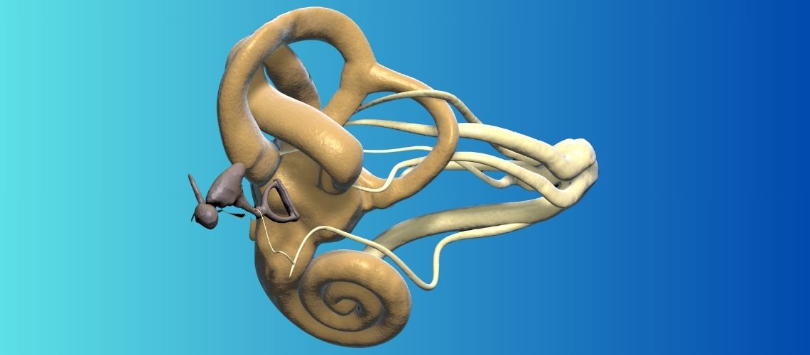
The command center of our body’s complex balance mechanism is the balance organ located in the inner ear. We can think of this area as a high-tech control room containing both motion sensors and gravity detectors. This control room has two main sections:
The first section consists of the semicircular canals. There are three fluid-filled canals in each ear, and they detect rotational movements of the head. Just as the air bubble inside a spirit level moves according to the tilt, the fluid inside these canals moves when we turn our heads. This fluid movement sends clear signals to the brain through the nerves, such as “we are turning right” or “we are bending down.” The most important feature of these canals is that under normal circumstances they are not affected by gravity. Their only task is to detect rotational movement.
The second section consists of two small sacs called the otolith organs. The structures popularly known as “ear crystals” are located inside these sacs. Scientifically called otoconia, these microscopic calcium carbonate crystals are embedded in a gelatinous layer. The function of these crystals is to act as tiny weights that detect gravity and linear movements (such as a car suddenly accelerating or an elevator moving upward). When we tilt our heads, these heavy crystals pull the gelatinous layer downward under the influence of gravity, providing the brain with information about the position of our head.
The problem begins when these crystals detach from their gelatinous layer and fall into a place where they do not belong, namely the semicircular canals. These canals, which normally respond only to fluid movement and are not influenced by gravity, suddenly become sensitive to gravity because of these dense and heavy crystals. As a result, when you move your head into certain positions, such as lying down in bed, these free-floating crystals begin to roll inside the canal like small pebbles. This rolling motion pushes the fluid inside the canal like a piston, sending a completely false signal to the brain that you are spinning violently even though you are actually still. This confusion of signals is what triggers that frightening vertigo attack.
What Causes Ear Crystals to Become Dislodged?
There may be more than one reason why the crystals detach from their normal location. Most of the time it is difficult to identify a single underlying cause, but certain conditions may trigger this process. The most common causes include:
- Advancing age (the most common cause)
- Blows to the head and/or neck
- Whiplash injuries following traffic accidents
- Sports injuries
- Previous ear disorders (Meniere’s disease, vestibular neuritis, vestibular migraine, etc.)
- Prolonged immobility after major surgical procedures
- Situations in which the head remains in the same position for a long time, such as sitting in a dental chair or a hairdresser’s chair
- Dehydration and lack of sleep
- Excessive salt or caffeine consumption
- Periods of intense stress
How Do These Dislodged Crystals Trigger a Vertigo Attack?
Two different mechanisms may occur depending on how the crystals behave inside the semicircular canal. Understanding which mechanism is responsible is critically important for selecting the correct treatment because each requires a different repositioning maneuver.
Canalithiasis (Free-Floating Crystals): This is the most common scenario and accounts for the vast majority of cases. The crystals move freely within the canal. When you place your head in the triggering position, it takes a few seconds before the crystals begin to move due to gravity. This explains why the dizziness starts after a short delay rather than immediately. Once the crystals settle at the lowest point in the canal, fluid movement stops, and the dizziness usually resolves spontaneously within less than a minute. This mechanism responds very well to repositioning maneuvers designed to return the crystals to their original location.
Cupulolithiasis (Adhered Crystals): This is a rarer and often more persistent condition. Here, instead of floating freely, the crystals adhere to the sensory organ (cupula) located at the end of the canal, much like a mussel attaching itself to a rock. This makes the normally neutral sensory organ heavier and transforms it into a constant gravity sensor. As soon as you move your head into the triggering position, the weighted cupula bends immediately, causing dizziness to begin instantly without any delay. As long as you maintain that position, the dizziness continues and may last much longer than one minute. Treating this condition may require different and more dynamic maneuvers to dislodge the attached crystals. Distinguishing between these two mechanisms is the most critical step in achieving rapid recovery.
Who Is at Greater Risk of Developing Ear Crystal Disorder (BPPV)?
Although this condition can occur in anyone, certain characteristics or existing health conditions may make a person more susceptible to BPPV. Knowing these risk factors is particularly important for people who experience frequent recurrences. These risk factors include:
- Being over the age of 50
- Sex (especially hormonal changes after menopause)
- Osteoporosis or osteopenia (bone loss or reduced bone density)
- Confirmed vitamin D deficiency
- A history of Meniere’s disease
- A diagnosis of vestibular migraine
- A previous head injury
- Uncontrolled high blood pressure
- Diabetes mellitus
The presence of these factors, especially those related to bone and calcium metabolism, demonstrates that overall body health directly affects inner ear health as well. Considering that these crystals are composed of calcium, it is not surprising that conditions affecting bone health may also impair the integrity of the crystals.
How Is the Definitive Diagnosis of Ear Crystal Displacement Made?
The diagnosis of BPPV is not established through imaging techniques such as MRI or CT scans. Instead, it is made entirely through the patient’s history and carefully performed positional assessments. Because the crystals are microscopic, they cannot be seen on any radiological examination. Accurate diagnosis is always achieved by observing, recording, and correctly interpreting the involuntary eye movements (medically known as nystagmus) that occur during provocative positional tests.
For this reason, the examination should be performed by audiologists specialized in this field. During the examination, Videonystagmography (VNG) or Frenzel goggles are generally used so that even the smallest eye movements can be clearly observed and to prevent the patient from unintentionally suppressing these movements by focusing on a fixed point.
The two most commonly used diagnostic tests are the Dix-Hallpike Test and the Supine Roll Test. The Dix-Hallpike Test is the gold standard for detecting crystals in the posterior (rear) and anterior (front) canals. In this test, while the patient is sitting upright, the head is turned 45 degrees to one side, and the patient is then quickly but carefully laid back into a supine position by the specialist. The Supine Roll Test is used when horizontal (lateral) canal involvement is suspected. During this test, the patient lies on their back while the head is turned to the right and left as eye movements are evaluated.
The direction, speed, onset time, and duration of the nystagmus observed during these tests clearly tell the specialist which ear is affected, which canal is involved, and which mechanism (free-floating or attached crystals) is responsible. This information is absolutely essential for selecting the correct repositioning maneuver.
Which Eye Movements Are Considered “Red Flags” That May Indicate a Dangerous Condition?
Some types of nystagmus observed during positional testing may indicate that the problem is not simple crystal displacement but rather a more serious neurological condition involving the brain or cerebellum. In such cases, urgent medical evaluation is required. These warning signs include:
- Severe nystagmus occurring even though the patient does not feel dizzy
- Pure upward- or downward-beating nystagmus without torsional movement when the head position changes
- Nystagmus that begins immediately when the head is moved and continues without fatigue as long as the position is maintained
- Eye movements that change direction even though the patient remains in the same position
- Associated symptoms such as double vision, speech impairment, difficulty swallowing, numbness, or weakness on one side of the body
How Is Ear Crystal Disorder Treated? Do Medications Work?
BPPV is not a disease that can be cured with medication because the problem is not chemical but entirely mechanical. Medications prescribed to suppress dizziness (antiemetics or vestibular suppressants) may reduce nausea and the sensation of spinning during the most severe hours of an attack, but they do not solve the underlying problem. This is similar to sitting on a softer seat to avoid feeling the bumps instead of repairing a flat tire.
The real and lasting treatment consists of a series of physical procedures called Canalith Repositioning Maneuvers (CRM), which are performed in a clinical setting. The goal of these maneuvers is to use gravity as an ally to carefully guide the misplaced crystals back to where they belong through a sequence of head and body movements. Once the crystals return to the otolith organ where they belong, they no longer trigger dizziness and are gradually dissolved and eliminated by the body.
What Are These Special “Ear Crystal Repositioning” Maneuvers Used in Treatment?
The specific maneuver used is determined according to the results of the diagnostic tests. Each canal and each mechanism has its own treatment pathway. The most commonly used maneuvers with the highest success rates include:
- Epley Maneuver
- Semont Maneuver
- Lempert Maneuver (also known as the Barbecue Roll)
- Gufoni Maneuver
Among these, the Epley Maneuver is the most commonly used treatment for the most frequently encountered posterior canal BPPV. It is so well known simply because posterior canal involvement is the most common form of the disorder. However, this does not mean that the Epley Maneuver is suitable for every patient. Therefore, these maneuvers should not be attempted at home.
The Semont Maneuver is another effective alternative for posterior canal involvement and involves rapidly moving the patient from one side to the other in a single motion. Although it may appear simple, the symptoms that occur during the maneuver must be carefully observed. For this reason, it should always be performed in a hospital setting by audiologists.
As its name suggests, the Lempert (Barbecue Roll) Maneuver requires the patient to be slowly rotated around their own body axis. This maneuver should only be used for lateral (horizontal) canal BPPV.
When these maneuvers are performed correctly, the success rate is very high, even after a single treatment session (80–90%). Therefore, having these maneuvers performed by a properly trained and experienced audiologist is critical for both the effectiveness and safety of treatment.
Is It Normal to Feel “Residual Dizziness” After Treatment?
Yes, this is a very common and completely normal condition. Even after successful repositioning maneuvers eliminate the severe vertigo attacks, many patients continue to experience mild dizziness, imbalance, a feeling of emptiness in the head, or the sensation of “walking as if drunk” for several days or even weeks. This condition is known as residual dizziness. The reason is that although the maneuver clears the canal, mild damage may still remain in the otolith organ (utricle), where the crystals originally detached, or the brain may still be adapting to normal signals after being exposed to abnormal signals for an extended period.
The best way to manage this condition is not to become fearful and remain inactive but to safely return to normal daily activities. Walking and gently turning the head from side to side help accelerate the brain’s recalibration process and allow the dizziness to resolve more quickly. Patients with residual dizziness benefit greatly from vestibular rehabilitation.
Why Does This Condition Recur and What Can Be Done to Prevent Ear Crystals from Becoming Dislodged Again?
The most frustrating aspect of BPPV is its high tendency to recur. Even after successful treatment, crystals may become dislodged again as long as the underlying predisposition that caused the first episode (such as age, bone health, trauma, etc.) remains. The annual recurrence risk is approximately 15–20%, and about half of all patients experience another episode within five years. However, this does not mean the condition is hopeless. Several steps may help reduce the risk of recurrence:
- First of all, it should be remembered that confirmed vitamin D deficiency is an important risk factor for recurrent BPPV. Individuals who experience repeated attacks are advised to consult a physician, have their vitamin D levels checked, and receive appropriate supplementation if a deficiency is present.
- Particularly in postmenopausal women, measures to protect bone health should be taken in light of the relationship between osteoporosis and BPPV.
- Keeping systemic diseases such as hypertension, diabetes, and high cholesterol under good control helps preserve inner ear circulation and indirectly supports crystal health.
- Patients should protect their heads from severe impacts and avoid sudden head movements whenever possible.
Frequently Asked Questions
Ear crystals most commonly become displaced due to age-related changes. In addition, head trauma, prolonged immobility, and certain inner ear disorders may cause the crystals to migrate into the semicircular canals.
Yes. As people age, natural changes occur in the supporting tissues of the inner ear, making it easier for the crystals to become displaced. For this reason, BPPV is seen more frequently in older adults.
Blows or concussions involving the head may cause the otolith crystals in the inner ear to detach from their normal position. If positional dizziness develops after trauma, an evaluation by an ear, nose, and throat specialist is recommended.
Prolonged immobility, postoperative bed rest, or remaining in the same position for extended periods may facilitate crystal displacement in some individuals and increase the risk of developing BPPV.
Meniere’s disease, vestibular neuritis, migraine, and certain inner ear disorders may increase the risk of crystal displacement. Identifying the underlying condition is important for treatment planning.
In addition to a detailed examination, diagnosis may include audiological evaluation, audiometry, audiograms, and when necessary vestibular tests such as VNG, vHIT, and VEMP. Positional tests also play an important role in diagnosis.
Although recurrence cannot be completely prevented, controlling underlying medical conditions, performing the recommended exercises, and attending regular follow-up appointments may help reduce the risk of recurrence.
In most patients, displaced ear crystals are treated successfully with repositioning maneuvers, and surgery is not required. Surgical treatment is considered only in very rare, resistant, and carefully selected cases.
If dizziness is accompanied by sudden hearing loss, speech impairment, double vision, altered consciousness, severe headache, or weakness in the arms or legs, immediate medical evaluation is necessary. These symptoms may indicate conditions other than BPPV.
If positional dizziness recurs, interferes with daily life, or is accompanied by hearing loss or tinnitus, you should consult an audiologist and an ear, nose, and throat specialist. Early evaluation allows appropriate treatment to be planned.

Dr. Audiologist Emel Uğur was born in 1982 in Çanakkale. She specialized in Pediatric Audiology, Otologic Disorders, and Vestibular System Disorders, and served for 15 years at the Istanbul Training and Research Hospital. In 2015, she joined the Acıbadem Healthcare Group. She currently works as an Audiologist at the Acıbadem Altunizade Hospital and also serves as a faculty member and program director in the Acıbadem University Vocational School of Health Services Audiometry Program.










Location of our clinic in Istanbul, Turkey