

 Türkçe
The acoustic reflex test is an audiological evaluation used to measure the neural response of the hearing system. This test records the reflex of the middle ear muscles to loud sounds and provides information about hearing health.
The question of how the acoustic reflex test is performed explains the application method. Sound is delivered through a probe placed in the ear canal, and the muscle response in the middle ear is measured. This method provides data about the auditory nerves and brainstem.
The importance of the acoustic reflex test becomes evident in determining the type of hearing loss. It is an effective diagnostic tool for distinguishing conductive, sensorineural, or central nervous system-related hearing problems.
The question of how acoustic reflex test results are interpreted requires expertise. The presence, absence, or threshold levels of the reflex provide information about the degree of hearing loss. This allows an appropriate treatment plan to be created.
What Is the Acoustic Reflex Test and Why Is It Important?
The Acoustic Reflex Test is a measurement method used to evaluate the automatic protective responses our ears produce against loud sounds. In other words, when our ear encounters an excessively loud stimulus, a small muscle located among the middle ear ossicles, known as the stapedius muscle, contracts reflexively and provides some protection to the inner ear against the potentially intense stimulus. This automatic reaction is a valuable clue showing how healthily our auditory system is functioning. The greatest importance of the test is that, although it is not a “hearing test” on its own, it reveals the effectiveness of the complex neural network extending from the ear canal to the brainstem. Not only the mobility of the eardrum or the function of the inner ear cells but also the transmission of the signal to the brain and then back to the muscles through feedback are important. Therefore, when this reflex mechanism cannot be activated or is triggered at abnormal levels, it becomes possible to gain insight into whether there is a problem in the ear or in the neural connections. For this reason, the Acoustic Reflex Test has become a subject of interest not only for healthcare professionals but also for individuals. By contributing to the early detection of certain sensory or neural conditions that may otherwise be difficult to identify, it opens the way to a healthier hearing journey. Especially in cases of one-sided complaints or suddenly developing hearing difficulties, the data obtained from this test play a key role in shortening the diagnostic process and ensuring appropriate referral.
How Is the Acoustic Reflex Test Performed?
This test can be evaluated with a specially designed device. In the first step, an appropriately sized earphone or probe is placed in the ear, and the ear canal is sealed so that no air leaks. This allows the device to establish pressure control capable of measuring the movement of the eardrum instantly. During the test, a fixed low-frequency tone is presented, and changes in the eardrum are recorded. Higher-intensity stimuli, meaning brief loud sounds, are then delivered to analyze whether the small muscle in the middle ear, the stapedius muscle, contracts. If the muscle contracts, the pressure and eardrum movement measured by the device change suddenly. The test is repeated for the main frequencies. This change is called the “acoustic reflex,” and the intensity of the recorded value, expressed in dB, indicates the threshold level of the reflex.
The person does not need to produce any special response during the test because the reflex occurs involuntarily. Therefore, it can also be easily performed on babies and children. Since the test generally takes no more than one or two minutes, it is a practical assessment method. In some cases, different stimuli may be delivered to both ears at the same time, or the test stimulus may be presented to one ear while the measurement is taken from the other ear. In this way, the reflex responses of both ears are evaluated comparatively. Through all these stages, the test reveals the integrity of the neural pathway extending from the outer ear to the reflex center in the brainstem and then back to the middle ear.
What Is the Physiological Basis of the Acoustic Reflex?
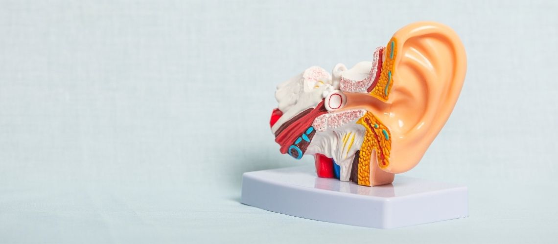
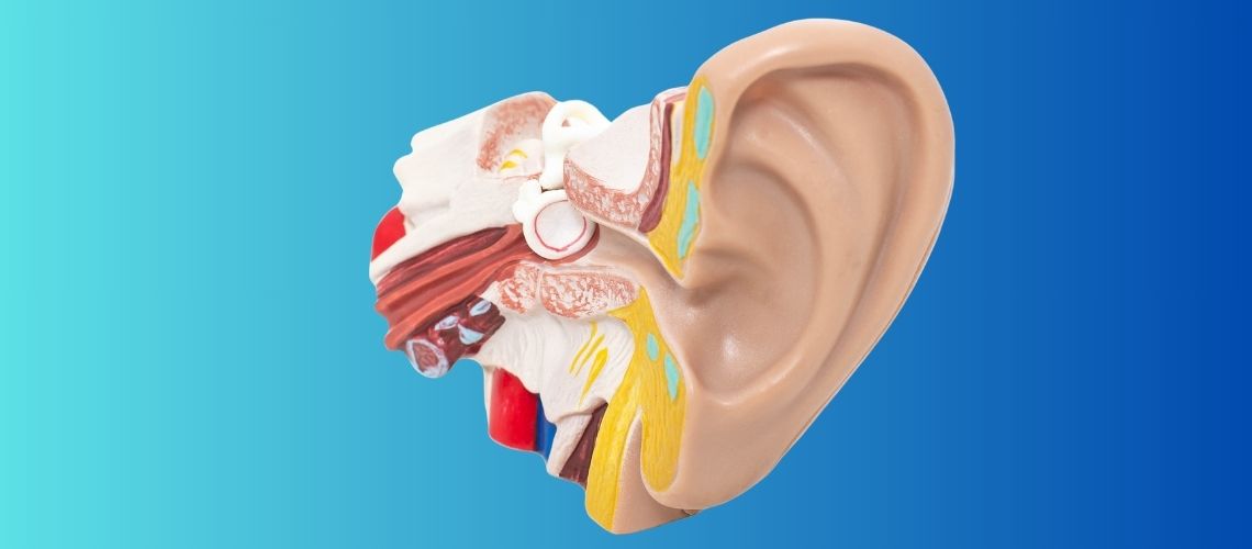
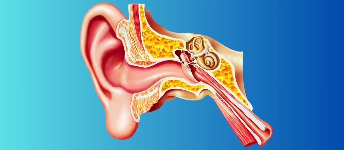
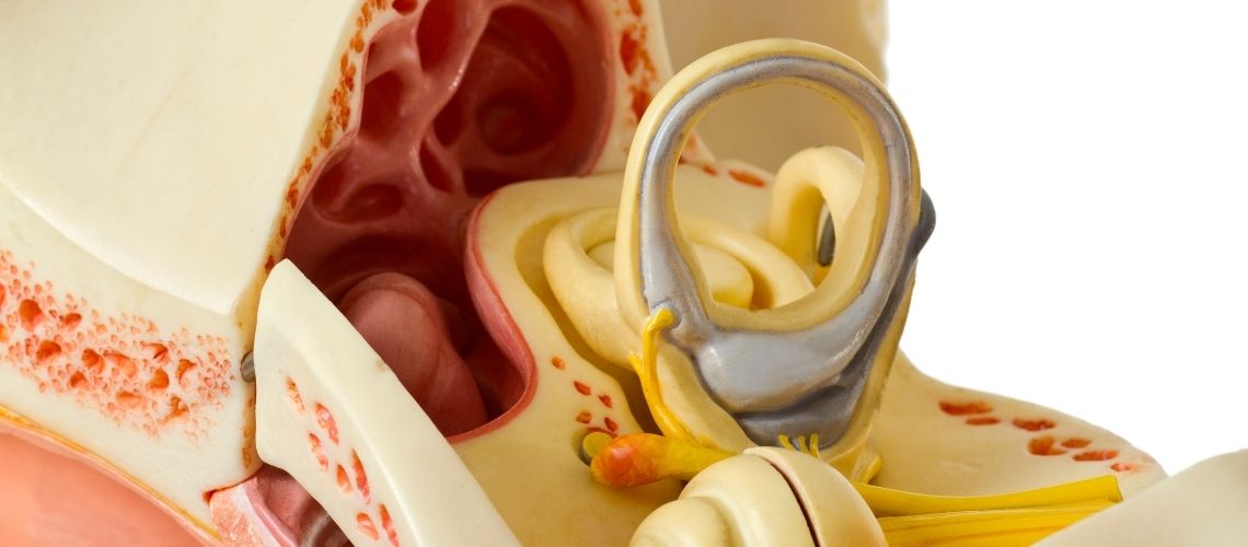
Like many automatic responses that develop in our body, the acoustic reflex is the result of a complex neural network. A high-intensity sound is transmitted from the outer ear to the middle ear and then activates the hearing receptors in the inner ear. The signals formed here travel along the auditory nerve and reach the auditory centers in the brainstem. When these signals are evaluated in the brainstem and a certain intensity threshold is exceeded, the brain’s feedback mechanism is activated. At this stage, a “contract” command is transmitted through the facial nerve to the stapedius muscle in the middle ear. When the stapedius muscle contracts, the pressure applied by the stapes to the oval window is slightly reduced, thereby decreasing the intensity of the sound transmitted to the inner ear to some extent. This means that our ear develops a type of defense mechanism when exposed to a sudden loud sound. Of course, this reflex cannot be considered a complete shield against extremely loud sounds, but it helps protect the inner ear structures by partially reducing low-frequency noise. There is an interesting interaction between the two ears because even a loud sound presented to only one ear may cause both stapedius muscles to contract simultaneously. This bilateral response provides more effective protection for the delicate structures of the ear. The key point is that the reflex is coordinated at the brainstem level and that smooth communication between the ear and the nervous system is essential.
Which Equipment Is Used During the Acoustic Reflex Test?
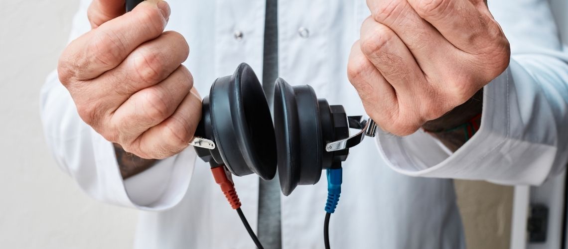
Special devices called tympanometers or immittance measurement instruments are mainly used to perform this test. The probe tip of the device generally contains a small speaker, a microphone, and a barometer that adjusts pressure. The speaker delivers a fixed-frequency, low-intensity tone to the ear. The microphone detects the reflected sound and pressure changes inside the ear. When necessary, a second earphone or insert may also be used to present a high-intensity test stimulus to the opposite ear or the same ear. Before the test begins, it is very important for the probe to fit the ear canal completely and be positioned without air leakage. Otherwise, the values obtained may be inaccurate. Modern devices largely automate the measurement process and display the results on a digital screen in the form of graphs or numerical values. In some systems, the pressure inside the ear is maintained close to zero while the probe is in place, and the reflex threshold measurement is performed in this position. The same device often also performs tympanometry, allowing additional information to be obtained about middle ear pressure balance and eardrum mobility. The patient generally does not need to make any special effort during use. A brief explanation is provided before the test, and the patient is informed that the sounds may seem somewhat loud. However, because the duration is short, the likelihood of discomfort is low. Since the current calibration of the equipment affects the accuracy of the results, regular maintenance and checks are performed.
How Are Acoustic Reflex Test Results Interpreted?
At the end of the test, it is recorded from which ear and at what intensity the reflex occurred or did not occur. These records are called threshold values and are generally expressed in dB. In individuals with normal hearing levels, the acoustic reflex is usually triggered within the range of 70 to 100 dB. If the reflex threshold is much higher than expected, it is reported as an “elevated” or “delayed” reflex. This may indicate a conductive or neural transmission problem in the auditory pathway. On the other hand, if the reflex does not occur at all, it is noted as “no reflex,” meaning “absent.” In this case, either the test sounds are not reaching the target structure or there is a significant interruption in neural transmission. For example, if there is fluid accumulation in the middle ear or a problem with the ossicles, the reflex may not occur. Similarly, if there is a problem in the auditory nerve or the reflex center in the brainstem, no response may be obtained during the test. For the result to be meaningful on its own, it is necessary to ensure that the individual has no additional pathology. This is because even earwax blockage may sometimes affect the measurement. Specialists evaluate acoustic reflex test results together with other hearing tests such as tympanometry. This provides a clearer idea of the level or structure at which the problem is located. Ipsilateral and contralateral measurements are also compared for each ear to obtain clues about the symmetry of the auditory system. This comprehensive approach facilitates rapid and accurate diagnosis and therefore treatment planning.
In Which Situations Is the Acoustic Reflex Test Used?
This test is frequently used to better understand the type and source of hearing loss. It provides supportive data, particularly when investigating whether there is a problem with the integrity of the eardrum or the ossicular chain. In conditions such as middle ear infection, fluid accumulation, or otosclerosis, the reflex generally does not occur at low intensity levels or cannot be recorded at all. Similarly, in the presence of progressive inner ear problems, reflex values may rise above the expected level. In people experiencing sudden hearing loss, checking acoustic reflex responses may also provide clues about the underlying cause. For example, if there is a one-sided hearing complaint and the reflex cannot be clearly measured in only one ear, a lesion in the auditory nerve or at the brainstem level may be suspected. In conditions such as tinnitus and hyperacusis, the test provides information for understanding the patient’s tolerance to loud sounds and observing potential protective responses within the ear. It may also be used as part of screening tests in newborn babies because babies can partially reveal their hearing function through involuntary reflex responses. Acoustic reflex data may also be used to obtain objective information in individuals with developmental delays or other communication difficulties. In this way, it becomes possible to identify the hearing status not only of adults but also of babies and children at an early stage and plan the necessary interventions in time.
What Should Be Considered During the Acoustic Reflex Test?
Before the test, it is important to ensure that the ear canal is clean and does not contain any foreign material causing obstruction. Even simple obstacles such as earwax may prevent the probe from fitting properly and make accurate measurement difficult. If a mild cold or upper respiratory tract infection is present, temporary changes in Eustachian tube function may cause differences in the test results. However, this generally does not constitute a serious problem. Excessive movement or activities such as chewing and swallowing during the test may reduce measurement accuracy. Sitting in a comfortable position and trying not to move the jaw whenever possible may be helpful. Being informed in advance that the sound stimuli may suddenly seem loud reduces the person’s anxiety. This is because head movements or involuntary reactions caused by nervousness may make recording the reflex more difficult. The test generally takes a very short time and does not cause pain or discomfort. If discomfort is felt during the test, the technician or staff member should be informed immediately. Some individuals are highly sensitive to loud sounds, and even brief stimuli may disturb them. In such situations, the test level may be kept within a range that the patient can tolerate. Finally, one of the most important factors affecting the test result is device calibration. Therefore, for reliable results, the selected clinic is expected to be meticulous and experienced in this regard.
Does the Acoustic Reflex Test Vary According to Age and Special Conditions?
As age advances, certain natural changes may occur in the ear structure and auditory pathways. Although the reflex threshold may rise slightly in middle-aged and older individuals, this does not always mean that hearing loss is present. Similarly, in babies and young children, measurement values may differ slightly because of anatomical differences in the ear canal. Especially during the newborn period, factors such as incomplete aeration of the middle ear or the eardrum not yet having reached its adult structure may affect test results. For this reason, higher-frequency probe tones may often be used in babies, allowing more accurate results to be obtained. The test may also be performed in individuals with special conditions, such as neurological developmental delay. In these individuals, the test is highly valuable because it provides objective data without requiring any behavioral response. Distinctive information may also be obtained from measurements in people who are candidates for cochlear implants or who use middle ear prostheses. This is because whether an acoustic reflex occurs in the ear where the prosthesis has been placed may provide insight into the success of the surgery. As a result, the ear structure of different age groups and special medical conditions may partially alter the acoustic reflex test protocol. However, the basic principle remains the same under all circumstances, and reliable results can be obtained in every age group when the test is performed correctly. In this way, many people, from babies to older adults, gain access to critical information about their hearing health.
Frequently Asked Questions

Dr. Audiologist Emel Uğur was born in 1982 in Çanakkale. She specialized in Pediatric Audiology, Otologic Disorders, and Vestibular System Disorders, and served for 15 years at the Istanbul Training and Research Hospital. In 2015, she joined the Acıbadem Healthcare Group. She currently works as an Audiologist at the Acıbadem Altunizade Hospital and also serves as a faculty member and program director in the Acıbadem University Vocational School of Health Services Audiometry Program.







Location of our clinic in Istanbul, Turkey