

 Türkçe
The eardrum is a thin and flexible structure located between the outer ear and the middle ear. Medically known as the “tympanic membrane,” it plays an important role in the hearing process by converting sound waves into vibrations. Tears or perforations in the membrane may cause hearing loss and infections.
The most common condition among eardrum disorders is tympanic membrane perforation. It generally occurs as a result of infection, sudden pressure changes, or trauma. If the perforation is small, it may heal on its own, but surgical intervention may be required in cases of extensive damage.
Middle ear infections may cause inflammation of the eardrum. Acute otitis media is common in children and causes ear pain, fever, and a feeling of pressure in the ear. Treatment is generally carried out with antibiotics and pain-relieving medications.
Examination of the eardrum is performed with a device called an otoscope. This examination evaluates the color, integrity, and mobility of the membrane. In cases of hearing loss, the hearing level is determined through audiological tests, and the treatment plan is created accordingly.
What Is the Eardrum?
The eardrum is a thin and flexible membrane located between the outer ear and the middle ear. In medicine, it is called the “tympanic membrane.” It converts sound waves into vibrations and transmits them to the middle ear, forming the first mechanical stage of hearing. It also protects the middle ear from external factors. If it tears, hearing loss, pain, and the risk of infection may occur. Depending on the size of the tear, treatment may involve spontaneous healing or surgery.
Where Is the Eardrum Located in Our Body and What Does It Look Like?
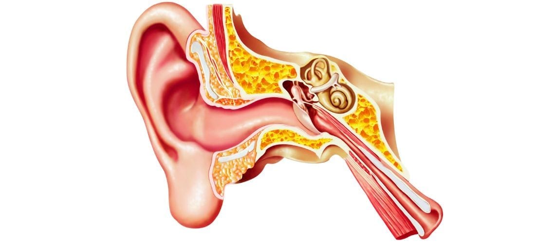
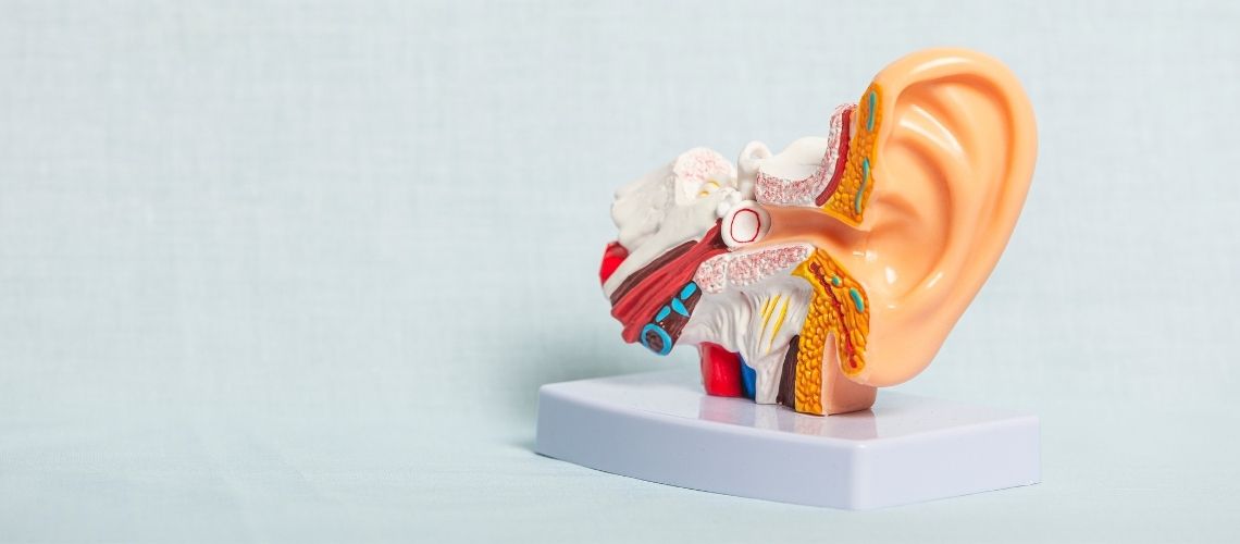
The eardrum, medically known as the tympanic membrane, is a curtain-like structure located at the very end of the external ear canal, immediately at the entrance to the middle ear cavity. It forms a delicate boundary between the outer ear and the middle ear.
A healthy eardrum has a very delicate structure. It is nearly transparent, slightly shiny, and has a pearly appearance. For this reason, it is described as “pearly gray.” It is approximately 1 centimeter in diameter, oval in shape, and contrary to common belief, it does not stand completely upright; it is slightly angled inward and forward. During examination, this angled position requires the physician to gently pull the auricle upward and backward in adults and slightly downward and backward in children in order to see it.
Although the eardrum appears to be a single piece, it actually consists of two main sections. We can compare these sections to the structure of a trampoline. You may think of it as the main tense surface on which we jump and the softer, more flexible edge band surrounding it.
One of the best instruments for visualizing the parts of the eardrum is a goblet drum. Just as the tight, firm upper skin called the “membrane” produces sound when struck on a goblet drum, it resembles the large, tense, and vibrating main section of the eardrum, known as the Pars Tensa. Like the firm upper surface of the drum, the eardrum is similarly the most important section that collects sound waves and vibrates to initiate the hearing process. The Pars Flaccida should be thought of as the part where the drum membrane connects to the rim or body. It is the smaller, looser, and more flexible section located at the edge of the membrane. This loose structure makes it more sensitive to pressure changes in the middle ear.
What Structural Features Make the Eardrum So Special?
The secret behind the eardrum being so thin yet so durable and functional lies in its three-layered “sandwich” structure. Each layer has a different function, and the harmonious operation of these layers is essential for perfect hearing. These three layers forming the eardrum are:
- Outer skin layer
- Middle fibrous layer
- Inner mucosal layer
The outer layer is a continuation of the skin lining the ear canal and has an incredible characteristic: it cleans itself. Cells continuously migrate outward from the center of the membrane, working like a moving staircase and carrying old cells out together with earwax. This mechanism keeps the eardrum clean and healthy.
The middle layer is the skeleton of the eardrum. It consists of specially arranged fibers that provide strength and flexibility. These fibers are woven like a spider web, both radially from the center outward and circularly. This cross-weaving allows the membrane to vibrate efficiently in response to sounds at every frequency while also giving it resistance to tension. The condition known as eardrum calcification, or tympanosclerosis, occurs when calcium accumulates in this middle layer and reduces the flexibility of the membrane.
The inner layer is an extension of the mucous membrane lining the middle ear cavity. This layer is like a mirror of the health condition of the middle ear. In conditions such as middle ear inflammation, this mucosa swells and thickens. One of the main reasons the eardrum appears red and dull during examination is inflammation of this inner layer.
How Do We Hear Sounds Through the Eardrum?
Hearing is a chain reaction that begins when sound waves are collected by the auricle, pass through the ear canal, and strike the eardrum. The eardrum acts as a converter, or transducer, that converts incoming air vibrations, or sound, into mechanical vibrations. Just like the diaphragm of a microphone, it vibrates with sound waves.
However, the true engineering marvel of the eardrum is that it performs a function called “impedance matching.” Our ear must transfer sound from air, a low-density medium, into the fluid of the inner ear, a high-density medium. You can imagine this by considering how difficult it is to hear someone calling to you from outside while you are underwater in a swimming pool. Most of the sound energy is reflected from the water surface. If there were no system in the middle ear to facilitate this transition, we would lose more than 99% of the sound and hear almost nothing.
The eardrum and middle ear ossicles solve this problem in two ways. The first is by creating a difference in area. The eardrum is much larger than the base of the stapes, through which sound is transmitted to the inner ear. By receiving the incoming force over a large area and concentrating it onto a very small point, it increases the pressure dozens of times. This is similar to how much pressure you apply to the ground when you place your entire body weight on the narrow tip of a high-heeled shoe. The second is that the eardrum and ossicles function like a lever system, further increasing this pressure. In this way, sound is efficiently transmitted to the fluid in the inner ear without losing energy.
How Does the Eardrum Protect Us from Loud Sounds and Noise?
The eardrum and middle ear system not only transmit sounds but also possess a natural defense mechanism that protects the inner ear from the harmful effects of loud sounds. This mechanism is called the “acoustic reflex” or “middle ear muscle reflex.” It functions like the body’s internal volume control button. When we encounter a loud sound, two tiny muscles in the middle ear contract involuntarily. This protective reflex has two main muscles and functions:
The stapedius muscle is activated in response to sudden and intense external sounds, such as an explosion or loud music. By contracting and stiffening the ossicular chain, it reduces the strength of sound vibrations reaching the inner ear. This provides particularly effective protection against low-frequency sounds.
The function of the tensor tympani muscle is more interesting. It reduces sound intensity by relaxing the eardrum during very loud sounds. This muscle is also activated mainly to suppress sounds produced by our own body, such as chewing, swallowing, and speaking. In this way, our internal noise does not prevent us from hearing important sounds coming from the outside world. If you have ever wondered why we do not hear the noise produced while chewing in an annoyingly loud way, the answer lies in the work of this small muscle. This muscle is also part of a general startle reflex.
How Is Otoscopy, the Examination of the Eardrum, Performed?
Examination of the eardrum, or otoscopy, is the most fundamental step in ear, nose, and throat and audiology practice. The small device with a light and magnifying lens at its tip that you see in your doctor’s hand is called an “otoscope.” This seemingly simple device provides highly valuable information about the health of the eardrum and external ear canal.
During the examination, the physician holds the otoscope like a pen while safely resting the edge of their hand against your cheek. This is an important safety maneuver that prevents the instrument from harming your ear even if you suddenly move. To see the eardrum clearly, the physician gently pulls the auricle upward and backward; this movement straightens the natural curve of the ear canal.
The main structures observed in a healthy eardrum are:
- Light reflection, light cone
- Head of the malleus
- Pearly gray color
- Transparency
- Correct position
Each of these findings has a meaning. For example, the bright triangular reflection known as the “light reflex” indicates the conical structure and health of the membrane. Its pearly gray and semi-transparent appearance suggests that there is no inflammation or fluid behind it. Being able to distinguish the malleus behind it is also a sign of a healthy membrane.
Sometimes your physician may perform an additional test called “pneumatic otoscopy.” This involves gently blowing air into the ear canal with the help of a small rubber bulb attached to the otoscope. This procedure tests the mobility of the eardrum. A healthy membrane should move slightly inward and outward with this air pressure. If the membrane does not move at all, this is usually one of the most important signs that fluid has accumulated behind it, indicating middle ear inflammation.
What Are the Common Problems That Can Be Seen in the Eardrum?
Otoscopy allows us to diagnose many ear conditions immediately. Looking at an eardrum is almost like taking an X-ray of the middle ear. Some of the most common pathological appearances observed during otoscopic examination are:
- Redness and bulging (Acute middle ear infection)
- Fluid accumulation and air bubbles (Otitis media with effusion)
- Perforation or tear (Perforation)
- White calcified spots (Tympanosclerosis)
- Inward collapse (Retraction)
- Soft tissue accumulation in the middle ear (Cholesteatoma)
These conditions are only a few of the pathological findings that may be observed during examination of the eardrum. After performing this examination, your physician may request various tests.
What Is a Tympanometry Test and What Information Does It Provide About the Eardrum?
Tympanometry is a rapid, painless, and highly valuable test that objectively measures the mechanical condition of the eardrum and middle ear system. It is not a hearing test; it can be considered more as a “pressure and flexibility check” of the middle ear. It is performed using a soft-tipped probe placed in your ear. While you hear a mild humming sound, the probe gently changes the pressure in your ear canal and measures how much your eardrum moves during this process. Three basic measurements are taken during this test.
- Ear canal volume
- Middle ear pressure
- Eardrum flexibility (mobility)
Ear canal volume tells us how large the space is between the tip of the probe and the eardrum. If this volume is much larger than expected, it means the eardrum is perforated because the device is now measuring not only the canal but also the middle ear cavity. Middle ear pressure is an indicator of how well your Eustachian tube is functioning. If the pressure is highly negative, this indicates that the Eustachian tube is not ventilating the middle ear sufficiently and that this may lead to fluid accumulation in the future. However, it is still not sufficient on its own. When Eustachian tube function needs to be evaluated, a specific Eustachian Function Test must be performed using the appropriate maneuvers. Eardrum flexibility shows how freely the membrane and the ossicular chain behind it can move. A very stiff system, meaning low flexibility, may indicate problems such as fluid or calcification in the middle ear, whereas an excessively flexible system suggests that there may be a disconnection in the ossicles.
How Are Tympanometry Result Graphs Interpreted?
A graph is obtained as a result of the tympanometry test. The shape of this graph provides clear information about the condition of the middle ear. Although it may appear complicated, there are essentially several main types. The most common types of results obtained from tympanometry and their meanings are as follows:
- Normal peak (Healthy middle ear) Type A
- Shallow peak (Stiffened middle ear) Type As
- Very high peak (Excessively flexible middle ear) Type Ad
- Flat line (Immobile eardrum) Type B
- Peak at negative pressure: Type C
- Peak at positive pressure: Type P
- Double normal peak: Type E (May develop due to previous infections)
A normal result appears like a sharp mountain peak directly in the center of the graph. This shows that middle ear pressure is normal and that the membrane has good flexibility. A shallow peak means that this mountain is flatter and lower, indicating that the system has become stiff, for example due to calcification. A very high peak, on the contrary, suggests that the system is excessively loose, for example due to ossicular discontinuity. A peak at negative pressure means that the mountain peak has shifted to the left side of the graph and is a typical finding of Eustachian tube problems.
One of the most important findings is the “flat line.” This means that there is no peak on the graph and indicates that the eardrum does not move at all. However, the cause of this condition cannot be understood on its own. This is where ear canal volume becomes important. If the volume is normal, a flat line means that there is fluid in the middle ear. If the volume is very large, this means that the membrane is perforated. If the volume is very small, it may simply indicate that the ear canal is blocked with earwax.
How Are Visual Examination and Tests Used Together in the Diagnosis of Eardrum Conditions?
In clinical practice, reaching the most accurate diagnosis depends not on a single finding but on bringing all the evidence together. Otoscopy, or visual examination, and tympanometry, or objective testing, work together like detectives to reveal the secrets of the eardrum and middle ear.
For example, let us consider a child who presents with complaints of fullness in the ear and reduced hearing. During examination with an otoscope, we observe that the eardrum is dull and yellowish and that there are air bubbles behind it. This suggests to your physician that there may be fluid in the middle ear. The tympanometry test performed immediately afterward produces a flat line and a normal ear canal volume. This objectively confirms our visual suspicion: fluid has accumulated in the child’s middle ear, indicating otitis media with effusion.
In another scenario, let us consider a patient who has received a blow to the ear. A clear perforation is seen on the membrane during otoscopy. The tympanometry test again produces a flat line, but this time the ear canal volume is much larger than normal. This shows that the device has passed through the perforation and measured the middle ear cavity as well, numerically confirming our visual finding.
This integrated approach eliminates uncertainty and guides us toward the most accurate diagnosis and therefore the most effective treatment plan. While visual examination shows “what is happening,” objective tests reveal “why it is happening” and “how significant it is” in numerical terms.
What Are the Responsibilities of the Audiologist in Eardrum Surgery?
In conditions requiring surgical intervention, such as an eardrum perforation, the role of the audiologist remains critically important before, during, and after the operation. When the decision for surgery is made and afterward, we as audiologists monitor and manage the patient’s auditory journey.
The main objectives of the preoperative audiological evaluation are:
- Determining the current hearing level
- Checking inner ear health
- Estimating the hearing improvement expected from the operation
This initial evaluation helps determine whether you are a suitable candidate for surgery and establishes a baseline for measuring the success achieved after the operation.
After the operation, the audiologist’s role is to objectively document the success of the surgical intervention. The following are evaluated during postoperative follow-up examinations.
- Objectively measuring improvement in hearing
- Checking the mechanical condition of the new eardrum
- Confirming the success of the operation
The hearing tests performed clearly reveal the amount of hearing improvement. Tympanometry shows whether the repaired new membrane is intact, flexible, and functioning properly. These data are the most concrete evidence of the functional success of the operation for both the surgeon and the patient.
What Does an Audiologist Do in the Follow-Up of Ear Tubes?
Audiologists also play an important role in the follow-up of patients who have ventilation tubes inserted into the ear due to persistent fluid accumulation in the middle ear or frequently recurring infections. The purpose of these tubes is to ventilate the middle ear, balance pressure, and prevent fluid accumulation. Our task is to ensure that the tube is performing its function properly. During regular follow-up visits, ENT physicians examine the following points.
- Whether the tube is in place (Visual examination)
- Whether the tube is blocked (Visual examination)
- Whether the tube is open and functioning
Whether hearing has returned to normal, through a hearing test, is an issue that should be examined by audiologists.
Frequently Asked Questions

Dr. Audiologist Emel Uğur was born in 1982 in Çanakkale. She specialized in Pediatric Audiology, Otologic Disorders, and Vestibular System Disorders, and served for 15 years at the Istanbul Training and Research Hospital. In 2015, she joined the Acıbadem Healthcare Group. She currently works as an Audiologist at the Acıbadem Altunizade Hospital and also serves as a faculty member and program director in the Acıbadem University Vocational School of Health Services Audiometry Program.







Location of our clinic in Istanbul, Turkey