

 Türkçe
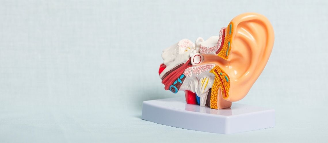
The outer ear is the anatomical structure that collects sound waves from the external environment and transmits them to the middle ear. It consists of two parts: the auricle (pinna) and the external auditory canal (meatus acusticus externus). It is the first line of defense and plays a critical role in the hearing process.
The auricle collects sound waves and directs them into the external auditory canal. The external auditory canal then transmits these waves to the eardrum. Earwax (cerumen) found within the canal traps foreign substances and protects against infections. Excessive cleaning may damage this protective structure.
The most common outer ear disease is inflammation of the external auditory canal (otitis externa). It usually develops due to swimming, trauma, or hygiene problems. It presents with symptoms such as ear pain, itching, discharge, and a sensation of blockage. Treatment is provided with drops and proper hygiene.
Tumoral formations may also rarely occur in the outer ear. In addition to benign masses, malignant lesions may also be present. If symptoms such as persistent itching, crusting, or bleeding occur, dermatological and ENT evaluation is important. Early diagnosis increases treatment success.
What Is the Outer Ear?
The outer ear is the first part of the ear that collects sounds from the external environment and transmits them to the middle ear. It consists of two main parts: the auricle (pinna) and the external auditory canal. It collects and directs sound waves toward the eardrum. It also keeps the ear canal moist and protected through the production of earwax (cerumen). This structure, which is the starting point of the hearing process, is also the ear’s first line of defense against infections.
Why Does Everyone’s Auricle Have a Different Shape and What Is the Role of This Shape in Hearing?
Like every person’s face, the auricle, medically called the pinna or auricle, is unique. This structure, which is as distinctive as a fingerprint, has vital functions that directly affect the quality of our hearing far beyond being an aesthetic feature. You can think of the auricle as a natural satellite dish or sound funnel designed to collect sounds as efficiently as possible.
Its basic framework consists of a single piece of yellow elastic cartilage that gives it both flexibility and a permanent shape. This cartilage is covered with a tight layer of skin that protects and nourishes it. The only part that does not contain cartilage is the earlobe (lobule), which consists of soft connective tissue and fat.
The complex and irregular surface of the auricle is not randomly formed. Each fold plays an important role in determining the direction and source of sound. In particular, our ability to understand whether a sound is coming from above or below is made possible by this unique structure. The folds of the auricle create tiny changes in the frequencies of sound waves before they reach our ears. Our brain interprets these subtle “acoustic signatures” and determines the position of sound on the vertical axis with remarkable accuracy. In addition, the forward-facing position of the auricles is a natural design that helps us distinguish whether sound is coming from the front or from behind.
The auricle has several main anatomical parts that help collect and direct sounds:
- Helix
- Antihelix
- Scaphoid Fossa
- Concha
- Tragus
- Antitragus
- Lobule (Earlobe)
In addition to these functions, the auricle also slightly increases sound intensity. In particular, the central hollow called the concha acts like a resonator and amplifies sounds around 4000–5000 Hz, one of the most critical frequency ranges of human speech, by approximately 10–15 decibels. It is a natural amplifier that helps us hear whispers or consonants in speech more clearly.
Why Is the Ear Canal Curved and What Is Its Structure Like?
The pathway that carries the sounds collected by the auricle to the eardrum is called the external auditory canal (external acoustic meatus). In an adult, this canal is approximately 2.5 cm long and is not a straight tube but has a slight “S” shape. This curved structure is actually an extremely intelligent natural defense mechanism. Just as the entrance to a castle is designed with turns to prevent intruders from entering directly, the S-shaped curve of the ear canal prevents foreign objects such as dust, dirt, and insects from easily reaching the sensitive eardrum.
For this reason, during an otoscopic ear examination, the auricle must be pulled upward and backward. This maneuver temporarily straightens the curved canal and allows the eardrum to be seen clearly.
The structure of the ear canal consists of two clinically important sections.
- Outer Cartilaginous Section: This forms the first one-third of the canal from the entrance and is a continuation of the cartilage of the auricle. The skin here is thicker and contains hair follicles, sebaceous glands that keep the skin moist, and special glands that produce earwax (cerumen). Most problems related to cotton swabs begin in this region.
- Inner Bony Section: This is the final two-thirds of the canal closest to the eardrum. This section is like a tunnel inside the skull bone (temporal bone). The skin covering it is extremely thin, sensitive, and directly attached to the underlying periosteum. There are no hairs or glands in this section. Therefore, this area is highly vulnerable to trauma and can easily bleed or become damaged even with minor intervention. The greatest danger of cotton swabs is that they push secretions from the outer part into this sensitive region, which cannot clean itself.
Almost all problems such as external ear inflammation (otitis externa) and earwax blockage occur in the outer cartilaginous section, where the glands and hairs are located.
Is Earwax (Cerumen) Really Dirt and Should It Be Cleaned?
Cerumen, commonly known as “earwax” and generally regarded as an unwanted substance, is actually a natural secretion that is vital for ear health. It is definitely not “dirt”; on the contrary, it is the ear canal’s personal protection and maintenance team. This substance is formed by the mixture of secretions from sebaceous and ceruminous glands with shed skin cells and performs many beneficial functions for our ears:
The benefits of earwax include:
- Moisturization: It keeps the canal skin moist and prevents dryness, itching, and cracking.
- Cleaning: It traps small particles such as dust, dirt, and pollen like an adhesive and prevents them from reaching the eardrum.
- Protection: Thanks to its water-repellent (hydrophobic) properties, it makes it more difficult for water to enter deeply and remain there during showering or swimming.
- Antibacterial and Antifungal Effect: Its mildly acidic structure, usually around pH 6.0, creates an environment unsuitable for the growth of bacteria and fungi.
Perhaps its most surprising feature is the ear’s self-cleaning mechanism. There is a skin migration process (epithelial migration) that begins at the center of the eardrum and slowly moves outward. You can think of this mechanism as a very slowly moving conveyor belt. This belt gradually carries old cerumen, trapped dust, and dirt toward the outside of the ear canal. At the end of this natural process, dried cerumen falls away unnoticed in small particles or accumulates in the auricle.
When cotton swabs are introduced at this point, the entire system is disrupted. Instead of cleaning the cerumen that the ear is trying to expel, these swabs push it deeper toward the sensitive bony canal, which cannot clean itself. This causes the cerumen to accumulate and harden there, forming earplugs called cerumen impaction. In short, the ear cleans itself perfectly; external intervention usually causes more harm than benefit.
How Does the Eardrum Transmit Sounds and How Can We Tell Whether It Is Healthy?
At the end of the external auditory canal lies the eardrum (tympanic membrane), a delicate structure that separates the outer ear from the middle ear. This membrane, which can be compared to the stretched skin of a drum or the diaphragm of a microphone, plays a critical role in the hearing process. Its primary function is to convert the acoustic energy created by sound waves traveling through the air into mechanical vibrations.
It has an oval, thin, and semi-transparent structure approximately 1 cm in diameter. It is not completely vertical but stands at a slight angle. This inclination helps it capture sound waves more efficiently. It consists of three different layers: an outer thin skin layer that is a continuation of the ear canal skin; a middle, regularly arranged fibrous layer that gives the membrane its strength and tension; and an inner mucosal layer that is a continuation of the mucous membrane lining the middle ear cavity.
During otoscopic examination (ear examination), a healthy eardrum generally appears pearly gray or slightly pinkish, transparent, and intact. One of the most important signs evaluated by the audiologist or physician during the examination is the triangular bright reflection created by the otoscope light in the lower anterior quadrant of the membrane. This is called the “cone of light.” A clear and bright reflection generally indicates that the eardrum is healthy, transparent, and in the correct position.
Other important structures seen in a healthy eardrum include:
- Pars Tensa: The large and tense section that forms the main vibrating part of the membrane.
- Manubrium: The handle of the malleus in the middle ear, which is attached to the inner surface of the membrane and appears externally as a whitish line.
- Umbo: The point where the lowest end of the malleus handle touches the membrane and where the membrane is most deeply retracted inward. The cone of light begins here.
- Pars Flaccida: A smaller and looser area located in the upper part of the membrane. It is weaker because it lacks the middle fibrous layer.
When the eardrum vibrates in response to sound waves, it transmits these vibrations directly to the ossicular chain in the middle ear (malleus, incus, and stapes), beginning the next stage of the hearing journey.
Why Do I Experience Ear Pain Even When There Is No Problem in My Ear?
This is a common and potentially confusing situation experienced by patients. You may feel severe pain in your ear, but when your doctor examines you, your auricle, canal, and eardrum may appear completely normal. The medical term for this condition is “referred otalgia,” meaning referred ear pain.
The reason is that the nervous system of the outer ear is highly complex and shares common nerve pathways with neighboring organs. Pain signals originating from a problem in one part of the body may be perceived by the brain as if they are coming from another location, in this case the ear. It is like crossed telephone lines: the source of the pain is different from the place where it is felt.
Possible sources of referred ear pain are quite varied.
- Temporomandibular Joint (TMJ) Problems: Conditions such as teeth grinding (bruxism), arthritis of the jaw joint, or disc displacement are among the most common causes.
- Dental Problems: Cavities, abscesses, or impacted wisdom teeth, especially in the back molars, may cause pain radiating to the ear.
- Tonsil and Throat Infections: Throat infections such as tonsillitis or pharyngitis frequently cause ear pain.
- Laryngeal and Pharyngeal Disorders: In more serious cases, a lesion or tumor in the larynx or pharynx may present first with referred ear pain.
- Neck Problems: A problem in the cervical vertebrae or even muscle spasm may be felt as pain in the ear.
Therefore, it is critically important for a person whose ear examination is normal but whose ear pain persists to consult an ENT physician or dentist to investigate the underlying cause. In addition, some people may develop a persistent cough reflex during examination or ear cleaning. This is also the result of the ear sharing common nerves with the throat and larynx and is completely normal.
Which Tests Does an Audiologist Use to Understand Outer Ear Problems?
The diagnosis of an outer ear problem includes a series of evaluations ranging from a simple visual examination (Otoscopic and Video Otoscopic Examination) to objective tests measuring the mechanical functions of the middle ear.
Everything begins with a visual examination performed using an otoscope, an instrument with a light and magnifying lens at its tip. This examination provides immediate information about the condition of the ear canal and eardrum. Today, this procedure is often performed using video otoscopes that project the image onto a screen. The greatest advantage of this technology is that you can also see the inside of your own ear on the screen. In this way, your ear, nose, and throat physician can allow you to see a problem being explained, such as cerumen blockage or inflammation, with your own eyes. This helps you understand the condition and increases your confidence in the treatment. In our country, this examination is performed by ENT physicians rather than audiologists. Audiologists perform otoscopic examinations only when necessary, such as during caloric testing or when taking an ear impression.
What Are Congenital Outer Ear Problems (Anomalies) and How Do They Affect Hearing?
Sometimes babies may be born with various developmental differences in the structures of the outer ear. These conditions may range from mild cosmetic problems to complex anomalies that seriously affect hearing.
Some of these anomalies include:
- Microtia: This means “small ear” and refers to the auricle being smaller than normal or incompletely developed. It has various degrees:
- Anotia: The congenital complete absence of the auricle.
- Aural Atresia: The congenital closure or complete absence of the external auditory canal. It is usually seen together with microtia.
- Canal Stenosis: The congenital narrowing of the ear canal. This condition may make a person more prone to earwax blockage and infections throughout life.
The most important consequence of these conditions, particularly in aural atresia, is maximum conductive hearing loss caused by sound being unable to reach the eardrum. This is a serious condition that may negatively affect the child’s speech and language development. However, the inner ears and auditory nerves of these children are generally healthy. Nevertheless, the child’s hearing must definitely be improved.
In such cases, conventional hearing aids cannot be used because there is no ear canal in which to place them. The solution is bone-conduction hearing systems that transmit sound directly to the healthy inner ear through the skull bone. In infants and young children, these devices are used without surgery by attaching them to an elastic band called a “softband.” Hearing provided by these devices at an early age enables the child to hear surrounding sounds and develop language skills alongside peers.
What Can Block the Ear Canal Other Than Cerumen Impaction?
Although cerumen impaction is the most common condition causing blockage in the ear canal, other structures may sometimes narrow or obstruct the canal. These are generally benign bony growths.
- Osteoma
Unlike exostoses, osteomas are true benign bone tumors that are usually solitary, pedunculated, and unilateral. They are less common.
Both conditions generally grow slowly and are considered for surgical removal (canalplasty) only when they cause symptoms such as blockage, recurrent infection, or hearing loss.
- Foreign Bodies
Especially in children, inserting objects such as small toy parts, beads, paper, or pieces of food into the ear canal is common. In adults, the tip of a cotton swab may occasionally break off and remain inside. These objects can easily be seen with an otoscope. Their removal, particularly when they are deep or located in a sensitive area, should generally be performed by an ENT specialist to prevent damage to the canal or eardrum.
Why Are Blows to the Ear Dangerous and When Is Emergency Intervention Required?
The unprotected position of the auricle on the body makes it highly vulnerable to trauma. Even a simple blow to the ear may sometimes cause serious and permanent consequences. The most urgent of these conditions is an auricular hematoma.
- Auricular Hematoma (“Cauliflower Ear”)
This condition is an otological emergency, particularly seen in contact sports such as wrestling and boxing. A hard, frictional blow to the ear causes the small blood vessels between the cartilage and the membrane surrounding and nourishing it (perichondrium) to rupture. As a result, blood accumulates between the cartilage and its membrane, causing a tense, painful, purple-red swelling in the auricle.
This is an emergency because the accumulated blood prevents the cartilage from receiving nourishment. If this blood collection is not urgently drained by a physician within 24–48 hours, the cartilage tissue dies due to lack of nourishment (necrosis). The body then attempts to repair this damaged area with irregular fibrous cartilage tissue. This process leads to permanent deformation of the auricle and the development of the well-known “cauliflower ear” deformity. When an audiologist or any healthcare professional identifies this condition, the patient must be referred immediately to an ENT specialist to prevent permanent damage.
Other traumatic injuries include cuts (lacerations) or partial or complete avulsion of the auricle. In such injuries, it is critically important that all exposed cartilage is properly covered with skin to prevent infection (perichondritis) and cartilage death, and medical intervention is essential.
Frequently Asked Questions

Dr. Audiologist Emel Uğur was born in 1982 in Çanakkale. She specialized in Pediatric Audiology, Otologic Disorders, and Vestibular System Disorders, and served for 15 years at the Istanbul Training and Research Hospital. In 2015, she joined the Acıbadem Healthcare Group. She currently works as an Audiologist at the Acıbadem Altunizade Hospital and also serves as a faculty member and program director in the Acıbadem University Vocational School of Health Services Audiometry Program.








Location of our clinic in Istanbul, Turkey