

 Türkçe
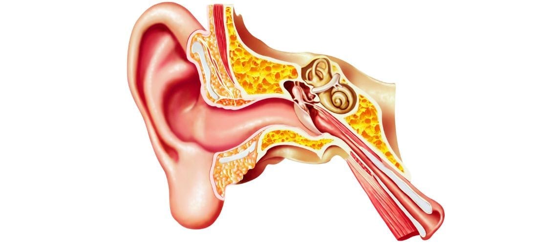
Hammer, anvil, and stirrup are the three tiny bones located in the middle ear that convert sound waves into mechanical vibrations during the hearing process. These bones are called the malleus (hammer), incus (anvil), and stapes (stirrup), respectively, and they transmit sound to the inner ear.
The malleus is directly attached to the eardrum and moves when the eardrum vibrates. This movement is transmitted to the stapes through the incus. The stapes then transfers these vibrations to the fluid of the inner ear through the oval window. This mechanical chain plays a critical role in sound transmission.
Hardening or damage affecting these bones may lead to conductive hearing loss. The most common example is otosclerosis, in which the stapes becomes fixed and vibration transmission is impaired. Surgical intervention, particularly stapedectomy, is among the treatment options.
Middle ear trauma, infections, or congenital anomalies may also affect the function of these ossicles. Diagnosis is made through otoscopy, audiometry, and temporal bone CT imaging. Structural abnormalities detected early can be corrected surgically, thereby preserving hearing function.
Where Are the Hammer, Anvil, and Stirrup Located in the Body and What Are Their Structures Like?
Holding the title of the smallest bones in the human body, the hammer (malleus), anvil (incus), and stirrup (stapes) are located in the middle ear cavity within the temporal bone of the skull. This cavity is like a miniature machine room, and these three ossicles are its most essential components. Connected to one another by delicate joints, they form an uninterrupted bridge extending from the eardrum to the inner ear, known as the ossicular chain. One of the most remarkable characteristics of these bones is that they complete their development in the womb and never grow again after birth.
Each link in the chain has its own unique structure and function:
The hammer, the outermost member of the chain, is attached directly to the eardrum. It resembles the tool from which it gets its name, and its “handle” is firmly connected to the inner surface of the eardrum. Thanks to this connection, even the slightest vibration produced in the eardrum by sound waves is instantly transmitted to the hammer. It is like touching the first domino; once the movement begins, it spreads throughout the entire chain. The head of the hammer forms a joint with the next bone, the anvil.
Located in the middle, the anvil serves as an intermediate station, transmitting the vibrations it receives from the hammer to the next link, the stirrup. Its projections extending in different directions allow it to transfer this mechanical energy as efficiently as possible. The long process where the anvil joins the stirrup is one of the most delicate parts of the chain because its blood supply is quite poor. For this reason, in conditions such as long-standing middle ear infections, bone erosion and damage are usually first observed in this region.
The final and innermost link of the chain, the stirrup, is truly an engineering marvel. As the smallest bone in the human body, it resembles a horse’s stirrup and rests on the oval window, the entrance to the inner ear. Its footplate is positioned like a piston that can move against this window. Its role is to transmit the mechanical vibrations received from the anvil to the fluid-filled cochlea of the inner ear. This final transfer of sound is one of the most critical steps in the hearing process. The stirrup is also the ossicle in which congenital structural differences and abnormalities are most commonly encountered.
The connections between these three ossicles are not simply attachments or fusions. Like our knees or elbows, they are connected by specialized synovial joints that allow movement. These flexible joints enable different movements depending on the frequency of the sound. As a result, they not only ensure the most efficient transmission of sound but also play an important role in the ear’s protective mechanisms.
Why Is the Sound Amplification Function of the Ossicular Chain So Important?
The primary purpose of the middle ear and its ossicular chain is to overcome a fundamental law of physics. Imagine trying to call out to a friend underwater. Your voice sounds muffled and weak because when sound passes from air into water, it loses a significant amount of its energy. Our external ear canal is filled with air, while our inner ear is filled with fluid. If sound waves attempted to pass directly from the eardrum into the fluid of the inner ear, more than 99% of the energy would be reflected back, resulting in a hearing loss of approximately 35 decibels (dB). This is almost the difference between a whisper and normal conversation.
The ossicular chain acts as a brilliant mechanical amplifier designed to prevent this major energy loss. It accomplishes this amplification through two basic principles:
The first and most effective is the area ratio principle. This principle works much like a thumbtack. A small force applied to its wide head creates enormous pressure at its pointed tip. The ossicular chain does the same thing. The surface area of the eardrum, where sound is collected, is approximately 20 times larger than the area of the stapes footplate, where sound is transmitted. By concentrating the sound energy gathered from the large surface of the eardrum onto the tiny base of the stapes, the ossicular chain dramatically increases the pressure. This hydraulic effect alone amplifies sound by approximately 26 dB.
The second principle is the lever effect. The ossicular chain functions like a lever or seesaw. The handle of the hammer is slightly longer than that of the anvil. This small difference in length creates a lever system that provides an additional but important increase in the force transmitted to the stapes. This lever effect contributes approximately another 2 dB of gain.
When these two mechanisms work together, they amplify sound by approximately 25 to 30 dB. This gain compensates for nearly all of the 35 dB loss that would otherwise occur when sound passes from air into fluid. As a result, even whispers from the outside world can be transformed into meaningful signals within the inner ear.
How Does the Middle Ear Protect Against Loud Sounds?
The role of the middle ear is not limited to transmitting and amplifying sound. It also has the vital responsibility of protecting the delicate hearing cells of the inner ear from potentially harmful loud sounds. This protection is provided through two different mechanisms: one active and one passive.
The Active Protection Mechanism is controlled by the middle ear muscles. The main player in this reflex is the stapedius muscle, which is attached to the stapes. When the ear is exposed to continuous loud sounds, generally above 70–100 dB, this muscle contracts automatically. This contraction stiffens the ossicular chain as a whole. The stiffened chain offers greater resistance to incoming sound vibrations and reduces the intensity of the sound transmitted to the inner ear by approximately 15–20 dB. This reflex has several important functions:
Reducing damage that may occur during prolonged exposure to loud sounds, such as at concerts or noisy workplaces.
Filtering low-frequency sounds such as rumbling and background noise, making speech easier to understand in noisy environments.
Reducing the intensity of our own body-generated sounds, such as speaking or chewing, so they do not become bothersome.
However, this reflex has a limitation: it works slowly. It cannot respond quickly enough to protect against sudden and extremely loud sounds such as gunshots or explosions.
This is where the Passive Protection Mechanism comes into play. This mechanism results from the natural flexibility of the joints between the ossicles. When a sudden and sharp sound wave, similar to an impact, arrives, these joints function as natural shock absorbers. They reduce the peak intensity of the incoming vibration, lowering its amplitude and transforming it into a softer, less harmful vibration. This protection is entirely mechanical and instantaneous.
The existence of this dual protection system has important implications, particularly in ear surgery. Modern prostheses used for ossicular chain reconstruction are generally rigid, one-piece structures. Although these prostheses transmit sound very effectively, they do not provide the passive shock-absorbing function of the natural joints. Therefore, a person whose hearing has been restored successfully through surgery should be aware that their inner ear may become slightly more vulnerable to sudden impact sounds than before.
What Are the Main Diseases Affecting the Ossicular Chain?
Unfortunately, this delicate and complex system can be disrupted by various diseases or trauma. These disorders prevent sound from being transmitted properly to the inner ear, leading to a condition known as conductive hearing loss. Problems affecting the ossicular chain can be grouped into three main categories.
Fixation (Hardening/Calcification): The chain loses its mobility, becomes rigid, and can no longer vibrate.
Discontinuity (Dislocation/Breakage): The connection between the ossicles is interrupted, or one of the bones erodes.
Erosion (Destruction): Usually caused by disease, resulting in gradual erosion and loss of the ossicles.
How Does Otosclerosis Affect the Ossicular Chain?
Otosclerosis, commonly known as “ear calcification,” is the most common cause of ossicular chain fixation. This disease is a unique form of abnormal bone remodeling affecting only the bony capsule surrounding the ear. As the disease progresses, abnormal spongy bone tissue typically develops around the oval window where the stapes footplate rests. Over time, this abnormal tissue hardens and effectively fuses the stapes to the oval window.
The stapes, which should function like a piston to transmit sound into the inner ear, becomes unable to move because of this fixation. This particularly impairs the transmission of low-frequency sounds. The result is a gradually progressive conductive hearing loss. It usually begins in individuals in their twenties or thirties, is more common in women, and often progresses during pregnancy.
Typical symptoms experienced by individuals with otosclerosis include:
- A slowly progressive condition that usually affects both ears (although it may also be unilateral). Audiometric evaluation typically reveals a characteristic conductive hearing loss known as Carhart’s notch at 2 kHz. If otosclerosis progresses and extends into the inner ear, it is referred to as cochlear otosclerosis.
- Persistent ringing in the ears (tinnitus).
- The sensation of understanding speech better in noisy environments (Paracusis Willisii). This occurs because the hearing loss filters low-frequency background noise, making speech sounds more prominent.
- Occasional episodes of dizziness.
What Causes Ossicular Chain Discontinuity?
Ossicular chain discontinuity, also known as ossicular dislocation, refers to the interruption of the mechanical connection between the ossicles. In this condition, sound cannot be transmitted effectively from the eardrum to the inner ear. This typically results in severe conductive hearing loss of approximately 50–60 dB, significantly affecting daily life. There are several possible causes of ossicular chain discontinuity.
- Head trauma or severe blows to the head, especially dislocation of the incudostapedial joint.
- Untreated or recurrent chronic middle ear infections.
- Cholesteatoma, which causes bone erosion within the middle ear.
- Previous ear surgeries involving the ossicles.
- As mentioned earlier, the most common discontinuity occurs when the long process of the incus, which has the poorest blood supply, erodes and loses its connection with the stapes.
What Is Cholesteatoma, a Dangerous Ear Disease?
Contrary to its name, cholesteatoma is neither a tumor nor cancer. Simply put, it is a condition in which “skin grows in the wrong place.” It develops when skin cells (epithelium), which normally line the external ear canal and the outer surface of the eardrum and constantly shed, somehow enter the middle ear cavity and accumulate there.
The middle ear has no mechanism for removing these skin debris. As a result, the debris accumulates layer upon layer like an onion, forming a cyst. What makes this cyst dangerous is that it releases enzymes capable of eroding bone as it grows. Cholesteatoma slowly but relentlessly destroys everything it comes into contact with. It first erodes the ossicles, causing hearing loss. If left untreated, it can erode the facial nerve canal, resulting in facial paralysis, destroy the inner ear leading to dizziness and complete hearing loss, and even erode the thin bony wall separating the middle ear from the brain, causing life-threatening complications such as meningitis or brain abscess. Because of its destructive nature, cholesteatoma must always be removed surgically once diagnosed. There are different types of cholesteatoma:
- Congenital Cholesteatoma: Arises from embryonic skin cells that remain in the middle ear during fetal development. It is usually seen as a white, pearl-like mass behind an intact eardrum.
- Primary (Acquired) Cholesteatoma: The most common type, usually resulting from chronic Eustachian tube dysfunction or untreated recurrent middle ear infections. Persistent negative pressure in the middle ear causes the weakest part of the eardrum to collapse inward, forming a pocket where skin debris accumulates and eventually develops into a cholesteatoma.
- Secondary Acquired Cholesteatoma: Develops when skin cells from the external ear canal migrate into the middle ear through an existing perforation in the eardrum.
The most common symptoms of cholesteatoma include:
- Long-standing ear discharge that is usually foul-smelling and may occasionally be bloody.
- Progressive hearing loss.
- A feeling of fullness or pressure in the ear.
- Dizziness.
- Facial paralysis on the affected side in advanced cases.
How Is the Diagnosis of Ossicular Chain Disorders Made?
When a patient presents with hearing loss, the diagnostic process resembles detective work. Audiologists and ear, nose, and throat specialists gather various clues to determine the underlying cause. This process ranges from a simple examination to advanced technological testing.
Everything begins with an otoscopic examination. Using a lighted instrument, the physician examines the ear canal and eardrum. Is the eardrum normal? Is there a perforation? Is there a visible mass or fluid behind it? These are the first clues. Next, pure tone audiometry is performed to determine the type and severity of hearing loss. During this test, the patient’s responses to sounds of different frequencies presented through both air and bone conduction are measured. In ossicular chain disorders, the typical finding is conductive hearing loss, where bone conduction hearing is normal or nearly normal while air conduction hearing is reduced. The difference between the two is called the Air-Bone Gap, and the size of this gap provides valuable information about the severity of the problem.
- An air-bone gap of 15–30 dB generally suggests middle ear fluid or an early-stage disorder.
- A gap of 30–40 dB may indicate a more serious ossicular disorder such as otosclerosis.
- A gap greater than 40 dB, particularly reaching 50–60 dB, is one of the strongest indicators of complete ossicular chain discontinuity.
Why Is Tympanometry Important in Diagnosis?
Tympanometry is an objective test that provides valuable information about the mechanical condition of the middle ear. It can be compared to checking the tension of a drum. Through a small probe placed in the external ear canal, gentle air pressure is applied to the eardrum while its flexibility is measured. The resulting graph, called a tympanogram, provides critical clues about the underlying condition.
- Type A: A normal tympanogram indicating normal middle ear pressure and mobility. However, this does not necessarily mean everything is normal. For example, a patient with an intact eardrum but an ossicular discontinuity may also have a Type A tympanogram.
- Type As: The “s” stands for stiffness. It indicates normal pressure but markedly reduced mobility and is a classic finding of ossicular fixation due to otosclerosis.
- Type Ad: The “d” stands for deep or discontinuity. It indicates normal pressure but excessively high mobility, which is a strong sign of ossicular discontinuity or an excessively flaccid eardrum.
- Type B: A flat tracing indicating no movement of the eardrum. The two most common causes are complete middle ear effusion or a perforated eardrum.
- Type C: Indicates that the peak occurs in the negative pressure range, providing direct evidence of Eustachian tube dysfunction and negative pressure within the middle ear.
What Is the Role of the Acoustic Reflex Test in Differential Diagnosis?
If the audiological test battery is a detective’s toolbox, the acoustic reflex test is the magnifying glass within that toolbox. It plays a crucial role in distinguishing between conditions that produce similar findings. This test measures whether the stapedius muscle contracts in response to a loud sound, indicating whether the ear’s natural protective reflex is functioning.
Imagine the following scenario: A patient has a 40 dB conductive hearing loss on audiometry and a Type A (normal) tympanogram. This pattern could be consistent with either early otosclerosis or ossicular discontinuity. This is where the acoustic reflex test becomes essential.
If the reflex is absent, it almost certainly indicates otosclerosis. Because the stapes is fixed by abnormal bone growth, the stapedius muscle cannot move the ossicular chain regardless of how strongly it contracts, making the reflex impossible to measure.
If the reflex is present (and the tympanogram is Type Ad), ossicular discontinuity should be suspected.
In this way, the acoustic reflex test is a highly valuable tool for distinguishing between different pathologies.
How Is Otosclerosis Treated Surgically?
The treatment for hearing loss caused by otosclerosis is surgical. The goal of the operation is to bypass the stapes, which has become immobilized due to abnormal bone growth. Today, the most commonly performed procedure is called a stapedotomy. During this operation, the surgeon works under a microscope to remove the movable upper portion of the stapes. A tiny opening, approximately the size of a pinhead, is then created in the fixed footplate using a laser or microdrill. Finally, a miniature piston prosthesis is placed between the incus and this opening to transmit sound.
Stapedotomy is one of the most successful procedures in otologic surgery. Its success rate exceeds 90%, and the vast majority of patients experience a significant improvement in hearing. However, like all surgical procedures, stapedotomy also carries potential risks. Your surgeon will inform you about these before the operation.
What Is Ossiculoplasty, the Surgical Treatment for Ossicular Chain Discontinuity?
Ossiculoplasty is the general term for surgery performed to reconstruct a disrupted or eroded ossicular chain. The aim of this operation is to restore the sound transmission pathway between the eardrum and the inner ear. The type of prosthesis used depends on the extent of the damage. If the upper part of the stapes remains intact, a PORP (Partial Ossicular Replacement Prosthesis) is used. If the stapes is completely absent, a longer prosthesis called a TORP (Total Ossicular Replacement Prosthesis) is required.
Many different materials have been used for this reconstruction over the years.
- The patient’s own cartilage or another intact ossicle (autograft).
- Titanium, a lightweight metal with excellent biocompatibility.
- Hydroxyapatite, a ceramic material with a structure similar to bone.
The results of ossiculoplasty are more variable than those of stapedotomy. Success depends largely on the overall health of the middle ear, particularly how well the Eustachian tube functions. Poor Eustachian tube function can lead to pressure problems in the middle ear after surgery, causing the prosthesis to become displaced or extruded by the eardrum. Therefore, the success rate of these procedures is approximately 60–70%.
Frequently Asked Questions

Dr. Audiologist Emel Uğur was born in 1982 in Çanakkale. She specialized in Pediatric Audiology, Otologic Disorders, and Vestibular System Disorders, and served for 15 years at the Istanbul Training and Research Hospital. In 2015, she joined the Acıbadem Healthcare Group. She currently works as an Audiologist at the Acıbadem Altunizade Hospital and also serves as a faculty member and program director in the Acıbadem University Vocational School of Health Services Audiometry Program.











Location of our clinic in Istanbul, Turkey