

 Türkçe
The Eustachian tube is a narrow canal located between the middle ear and the nasopharynx that equalizes ear pressure. Known medically as the tuba auditiva, it opens during swallowing or yawning, equalizes the air pressure in the middle ear with the external environment, and ensures the healthy functioning of the eardrum.
Eustachian tube dysfunction develops when this canal does not open sufficiently. In this case, complaints such as ear blockage, a feeling of pressure, hearing loss, and sometimes pain may occur. Allergies, infections, or anatomical narrowing may cause this condition.
Since the Eustachian tube is shorter and positioned more horizontally in children, susceptibility to middle ear infections (otitis media) increases. For this reason, ventilation tube placement may often be required in frequently recurring infections. Early intervention prevents permanent hearing loss.
Diagnosis is made through tympanometry and endoscopic examination. Nasal decongestant sprays, antihistamines, and surgical methods in appropriate cases are used in treatment. In chronic cases, procedures such as tube placement or adenoid removal may be required.
What Is the Eustachian Tube?
The Eustachian tube is a canal approximately 3–4 cm long that connects the middle ear to the nasopharyngeal cavity. Its primary function is to equalize the air pressure in the middle ear and prevent fluid accumulation. It opens during swallowing, yawning, or chewing to maintain this pressure balance. When blocked, a feeling of fullness in the ear, hearing loss, tinnitus, or infection may occur. Treatment is planned according to the underlying cause.
What Does the Eustachian Tube, This Small Canal in Our Body, Do?
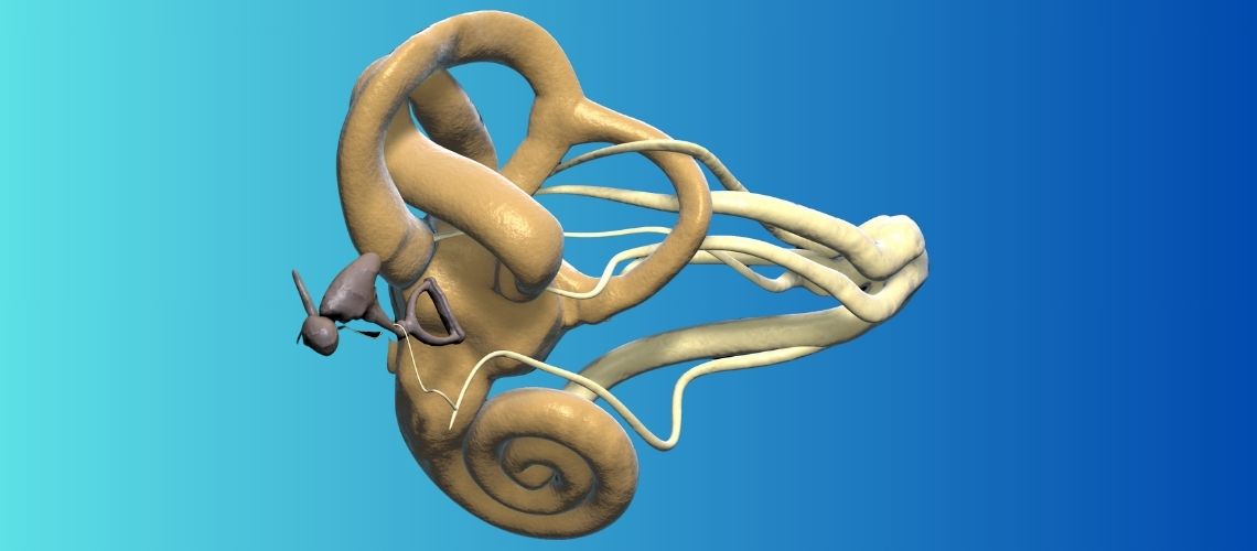
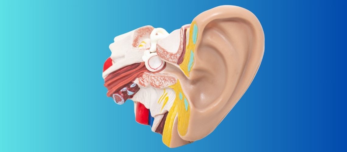
The middle ear is an air-filled chamber located immediately behind the eardrum and contains the tiny bones we call the malleus, incus, and stapes. For sound to be transmitted correctly to the inner ear, the air pressure inside this chamber must be the same as the atmospheric pressure outside. The structure that maintains this critical balance is the Eustachian tube, which is approximately 3.5 centimeters long.
This tube starts from the middle ear, extends downward, forward, and toward the center at a slight angle, and opens into the nasopharynx. Its structure is quite special; the one-third section closest to the ear consists of bone, while the remaining two-thirds section closest to the nasopharynx consists of cartilage and soft tissue. This cartilaginous section normally remains closed like a valve. When we swallow, yawn, or chew something, certain specialized muscles around the tube contract and momentarily open this valve. During this brief opening, fresh air enters the middle ear, the pressure inside is equalized with the outside, and any accumulated fluids are drained toward the nasopharynx.
In summary, the Eustachian tube has three fundamental and vital functions in our body:
Pressure Equalization (Ventilation): This is perhaps the function we feel most clearly. The air in the middle ear is continuously absorbed by the surrounding tissues. This creates slight negative pressure inside, in other words a vacuum effect. The Eustachian tube opens hundreds of times a day, eliminates this pressure difference, and ventilates the middle ear. The “pop” sound we feel in our ears while an airplane is descending is actually a sign that our Eustachian tube has successfully performed its pressure-equalizing function.
Cleaning (Drainage): The middle ear is a self-cleaning system. The inner surface of the Eustachian tube is covered with tiny hairs (cilia) that move in one direction toward the nasopharynx and mucus-producing cells. This structure carries unwanted fluids, inflammatory residue, and microbes that may form in the middle ear toward the nasopharynx like an escalator. In this way, it helps keep our middle ear sterile and protects it from infections.
Protection: By normally remaining closed, the Eustachian tube acts as a shield that protects our middle ear from microbes, secretions, and even acidic contents escaping upward from the stomach that may be present in the nasopharynx and cause infection. It also prevents body sounds such as our own breathing or speaking voice from traveling directly to the middle ear and being heard in a disturbing manner.
Any disruption in these functions leads to a range of problems that we call “Eustachian tube dysfunction” and that directly threaten ear health.
So, Why Does the Eustachian Tube Become Blocked or Lose Its Function?
The failure of the Eustachian tube to perform its function most commonly occurs as a result of swelling and inflammation of the membrane lining the inside of the tube (mucosa). Just as we have difficulty breathing when our nose is blocked, when the inside of the Eustachian tube swells, it becomes unable to perform its normal opening and closing movement. Many factors may lead to this condition.
Some of these factors are as follows:
- Upper respiratory tract infections such as the common cold and flu
- Allergic rhinitis (hay fever)
- Chronic sinusitis
- Exposure to cigarette smoke
- Laryngopharyngeal reflux (silent reflux)
- Gastroesophageal reflux (acid reflux)
- Adenoid enlargement (especially in children)
- Tumors in the nasopharyngeal region (rarely)
- Congenital anomalies such as cleft palate
- Down syndrome
- Muscle dysfunction or loss (such as frequent weight gain and loss)
- Teeth grinding at night
These factors cause the tube either to become physically blocked or to fail functionally, laying the groundwork for ear problems. Especially in unilateral and long-lasting ear blockage that develops in adults, a detailed examination of the nasopharyngeal region by a physician is of great importance in ruling out more serious underlying causes.
Which Symptoms Indicate Eustachian Tube Problems?
Eustachian tube dysfunction is not a single disease; it is a collection of problems that may appear in different forms depending on the underlying cause. In clinical practice, grouping these problems into three main categories is critically important for making the correct diagnosis and planning the most effective treatment. This is because the treatment of each group is not only different but may sometimes be completely opposite.
- Obstructive (Dilation Disorder) Eustachian Dysfunction: When the Tube Cannot Open Sufficiently
This is the most common type of Eustachian tube problem. The primary issue is that the tube cannot open as it should. The inside of the tube usually swells after an infection or allergy, and the valve mechanism effectively becomes “stuck.” When the tube cannot open, the air in the middle ear continues to be absorbed, creating a vacuum effect inside. This negative pressure then pulls the eardrum inward. The symptoms caused by this condition are generally as follows:
- A feeling of fullness or pressure in the ear
- The ear feeling “blocked” or “filled with water”
- Muffled or reduced hearing
- Crackling, clicking, or popping sounds from the ear while swallowing or yawning
- Ear pain felt especially during pressure changes
- Tinnitus or humming
If this blockage persists for a long time, fluid begins to leak from the body into the middle ear cavity due to the effect of the negative pressure. This condition is medically called otitis media with effusion (OME) and is popularly known as fluid accumulation in the middle ear. This is one of the leading causes of hearing loss, particularly in children, that may negatively affect language and speech development when it goes unnoticed.
- Patulous (Open) Eustachian Dysfunction: When the Tube Cannot Close as It Should
This is a less common problem that is the exact opposite of obstructive dysfunction. The problem here is that the Eustachian tube remains continuously open when it should close. This causes uncontrolled airflow between the middle ear and the nasopharynx. The most common cause is rapid and significant weight loss. With weight loss, the fatty tissue around the Eustachian tube that normally helps it remain closed disappears, and the tube loses support and remains abnormally open.
The most disturbing and typical symptom of a patulous Eustachian tube is autophony. This is the condition in which a person hears their own voice and even their breathing sounds (tympanophony) abnormally loudly, with an echo, and “as though speaking from inside a barrel.” This condition may be extremely disturbing for patients. A patulous Eustachian tube also has other characteristic symptoms:
- Hearing one’s own voice or breathing in the ear
- A feeling of fullness in the ear
- Reduction of symptoms when lying down or bending the head forward
- A constant urge to sniff
Patients generally state that they feel relief when lying down or bending their heads forward because blood circulation in the head region increases in these positions, temporarily helping the tube close.
- Baro-Challenge-Induced Eustachian Dysfunction: When It Cannot Withstand Sudden Pressure
This is a situational functional disorder. The Eustachian tubes of these patients function without problems in normal daily life. However, during sudden and severe pressure changes such as air travel, especially during descent, scuba diving, or rapidly changing altitude on a mountainous road, they cannot open quickly enough to equalize pressure. The high pressure in the external environment can effectively “lock” the tube inward, and as the tube remains unable to open, the pressure difference increases further, creating a vicious cycle. The symptoms of this condition occur only during such barometric challenges.
- Sudden onset of severe ear pain
- An intense feeling of pressure in the ear
- Sudden muffling of hearing
- Sometimes dizziness
The ear examinations and audiological tests of these patients performed under normal conditions generally produce completely normal results. Diagnosis is largely based on the patient’s typical history.
How Are Eustachian Tube Problems Diagnosed?
Diagnosing Eustachian tube dysfunction is similar to putting together the pieces of a puzzle. The process begins by carefully listening to the patient’s description, continues with a physical examination, and is completed, most importantly, with audiological tests that objectively measure function. The aim is not merely to say, “There is a Eustachian tube problem,” but also to clearly determine which type of problem exists, whether obstructive, open, or situational. This is because the correct diagnosis is the first and most important step toward the correct treatment.
First Steps: Medical History and Examination
- Patient History: This forms the basis of diagnosis. It is highly valuable for the patient to describe the complaints in detail, when they began, what triggers them, for example whether they began after the flu or after air travel, and what provides relief, for example whether they resolve when lying down. Distinguishing between the feeling of “fullness” in obstructive dysfunction and true “autophony” in a patulous Eustachian tube, meaning hearing one’s own voice, is a critical clue on the path to the correct diagnosis.
- Physical Examination (Otoscopy): This involves carefully examining the ear canal and eardrum with the help of an otoscope. In obstructive dysfunction, the eardrum may appear retracted inward, its color may appear dull, or a yellowish color or air bubbles indicating the presence of fluid in the middle ear may be seen. In a patulous Eustachian tube, it may sometimes be observed that the eardrum moves slightly inward and outward in synchronization with the patient’s breathing. This is an important finding that supports the diagnosis.
Second Step: Objective Evidence Through Audiological Evaluation
Although the patient’s descriptions and examination findings provide direction, objective tests are needed to establish a definitive diagnosis and determine the degree of the problem. At this point, audiology comes into play and reveals the function of the Eustachian tube and its effects on the middle ear through concrete data.
- Pure Tone Audiometry (Hearing Test): This test is performed to determine the presence, type, and degree of possible hearing loss. Obstructive Eustachian dysfunction, particularly when accompanied by fluid accumulation in the middle ear, typically causes conductive hearing loss because it prevents sound from being transmitted to the inner ear. This loss is usually more pronounced at low frequencies.
- Tympanometry: This test is the gold standard for evaluating Eustachian tube function. It is a painless and very brief test that objectively measures middle ear pressure and eardrum mobility. Through a small, soft-tipped probe placed in the ear, slight air pressure is applied to the external ear canal, and the eardrum’s response to this pressure is plotted as a graph. This graph is called a tympanogram and provides us with highly valuable information.
Type A Tympanogram: This is a normal, bell-shaped curve. It indicates that middle ear pressure is normal. We see this result in healthy individuals, in a patulous Eustachian tube, and in baro-challenge dysfunction while at rest.
Type C Tympanogram: This is a result in which the peak of the curve shifts toward negative pressure. It is a classic indicator that negative pressure has accumulated in the middle ear and that the Eustachian tube is not performing its ventilation function effectively. It is a typical finding in obstructive dysfunction.
Type B Tympanogram: This appears as a flat line. It indicates that the eardrum does not move at all. The most common cause is fluid accumulation in the middle ear as a result of chronic obstructive dysfunction (otitis media with effusion).
Third Step: Advanced Function Tests When Necessary
Standard tympanometry captures a snapshot of the current condition and provides information about the consequences of Eustachian tube problems, such as negative pressure or fluid. However, sometimes, especially when standard tests are normal but the patient’s complaints persist, we need to test the Eustachian tube itself dynamically, in other words “while it is working.” Advanced function tests serve exactly this purpose.
Standard Eustachian Function Test: First, tympanometry is performed, followed by two additional tympanometry measurements after the Toynbee and Valsalva maneuvers to determine whether the necessary pressure change has occurred. The amounts of pressure change are evaluated.
Nine-Step Test (Bluestone Method): This classic test measures how effectively the Eustachian tube equalizes artificially created pressure through swallowing. Using a tympanometry device, positive and then negative pressure is first applied to the ear, and the patient is asked to swallow each time. The improvement in middle ear pressure after swallowing is evidence that the tube is actively functioning. If no pressure change occurs, this indicates an obstructive problem.
Sonotubometry: This test allows us to directly detect through sound waves whether the Eustachian tube opens. During the test, a sound source is placed in one nostril and a sensitive microphone is placed in the ear canal on the same side. The patient is asked to swallow. If the Eustachian tube opens during swallowing, the sound in the nasopharynx passes through the middle ear and is detected by the microphone in the ear canal. This appears as a sudden increase in sound on the test recording and provides clear evidence that the tube has opened.
Tubomanometry (TMM): This modern technique measures the efficiency of the Eustachian tube, in other words how quickly it can open under controlled pressure. As though simulating the descent of an airplane, controlled pressure is applied to the nasopharynx while the patient swallows, and at the same time a sensor in the ear canal precisely measures how long it takes for this pressure to reach the middle ear. The results show whether the tube opens on time or with a delay. Delayed opening indicates obstructive (dilation disorder) dysfunction.
Which Treatment Methods Are Used for Eustachian Tube Problems?
The treatment of Eustachian tube dysfunction does not consist of a single solution like the touch of a magic wand. The treatment plan is individually shaped according to the type of problem, whether obstructive or open, the underlying cause, whether allergy or infection, and its severity. Treatment generally begins with the simplest methods requiring the least intervention, and if these are insufficient, the next step is taken.
First Step: Conservative and Medical Treatments
In obstructive Eustachian dysfunction, our first goal is to reopen the tube and reduce the swelling (edema) inside it.
- Physical Maneuvers (Autoinflation): These are first-line methods that the patient can apply independently and that aim to mechanically open the tube.
Valsalva Maneuver (Trying to exhale while the mouth and nose are closed)
Toynbee Maneuver (Swallowing while the nose is closed)
Swallowing frequently
Yawning
Chewing gum
Medical devices such as Otovent®, in which a special balloon is inflated through the nose
- Medication Treatment: These treatments generally target the underlying inflammatory cause.
Why Is the Eustachian Tube Different in Children Than in Adults?
Ear problems in children, particularly those involving the middle ear, are both more common and have different characteristics compared with adults. The basis of this condition lies in the anatomical and physiological differences present during children’s development.
Anatomical Differences:
A child’s Eustachian tube is structurally more disadvantaged than that of an adult.
It is shorter.
It is wider.
It lies at a more horizontal angle, compared with the approximately 35-degree steep angle in adults, while this angle is only around 10 degrees in children.
This flatter position prevents gravity from helping drain fluid from the middle ear toward the nasopharynx. At the same time, it makes it much easier for microbes and secretions in the nasopharynx to flow backward into the middle ear (reflux). In addition, the muscles and cartilaginous structure that open and close the tube have not yet fully developed. This causes the tube to function more “loosely” and less efficiently. As the child grows, by approximately 7–8 years of age, the Eustachian tube lengthens, its angle becomes steeper, and it reaches its adult form. This is also the reason why the frequency of middle ear infections decreases significantly after these ages.
Different Causes and Consequences:
The most common causes underlying Eustachian tube blockage in children are also different from those in adults.
Adenoid Enlargement (Adenoid Hypertrophy): The adenoid is lymphoid tissue located immediately next to the opening of the Eustachian tube into the nasopharynx. The physiological enlargement of this tissue during childhood is the most common factor that mechanically blocks the opening of the tube and causes obstruction.
Frequent Upper Respiratory Tract Infections: Children’s developing immune systems and their presence in shared environments such as nurseries and schools make them more susceptible to viral infections that block the Eustachian tube.
For these reasons, Eustachian tube dysfunction in children often results in acute otitis media (sudden middle ear infection) and otitis media with effusion (fluid accumulation in the middle ear). In a child who cannot yet speak, the symptoms of this condition may include more general signs such as:
- Restlessness
- Fever
- Sleep disturbance
- Pulling or scratching the ear
The main concern is the potential for fluctuating hearing loss caused by fluid accumulation in the middle ear to negatively affect the child’s speech and language development during the most critical learning period. For this reason, the treatment approach in children may generally be more proactive and surgically oriented than in adults. In cases of recurrent middle ear infections or chronic fluid lasting longer than 3 months and affecting hearing, ear tube placement and/or adenoid surgery are frequently preferred treatment methods to protect development.
Frequently Asked Questions

Dr. Audiologist Emel Uğur was born in 1982 in Çanakkale. She specialized in Pediatric Audiology, Otologic Disorders, and Vestibular System Disorders, and served for 15 years at the Istanbul Training and Research Hospital. In 2015, she joined the Acıbadem Healthcare Group. She currently works as an Audiologist at the Acıbadem Altunizade Hospital and also serves as a faculty member and program director in the Acıbadem University Vocational School of Health Services Audiometry Program.









Location of our clinic in Istanbul, Turkey